Anti-A antibodies in plasma. Note the position of the hands palms forward with thumbs outward and the feet toes pointed.

A P Chapter 1 Quiz Flashcards Quizlet
Before you begin the activity print out this graph paper so your students can draw the letters of their name using clean straight lines.

. By Andrea Kirsh November 16 2021. B antigens on cell. The blood flow through the ascending and descending portions of the vasa recta.
Anti-B antibodies in plasma. Identify the functional area of the kidney at letter B. Moving from simpler to more complex which level of organization is immediately before simpler than the cell.
ICT-8-11 - Future Networks D14 Simulations and physical layer validations. In this study cyclic Arg-Gly-Asp-d-Tyr-Lys cRGDyK was conjugated with 2- p -isothiocyanatobenzyl-147-triazacyclononane-147-triacetic acid SCN-Bz-NOTA and then labeled with 68Ga. Selecting or hovering over a box will highlight each area in the diagram.
Use the same blank photos. Figure 23 Blood Type. Percentage of total mobile data traffic to be offloaded by 2018.
A brand is a name picture design or symbol or combination of those items used by a seller to identify its offerings and to differentiate them from competitors offerings. Body cavities are spaces within the body that surround internal organs. Figure 45 Label the stages of cell division and interphase.
Flowchart of the Major Arteries of the Trunk 2 of 3 Part A Drag the labels to the appropriate location in the figure. Branding is the set of activities designed to create a brand and position it in the minds of consumers. Show transcribed image text Expert Answer.
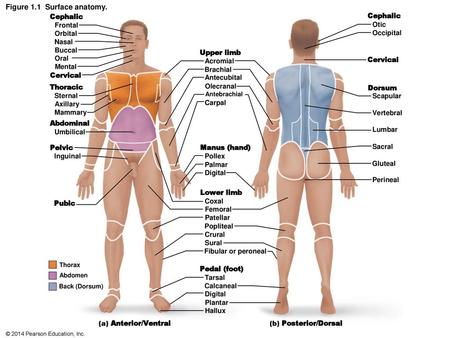
The individual in this figure is in anatomical position. Arg-Gly-Asp RGD derivatives have been labeled with various radioisotopes for the imaging of angiogenesis in ischemic tissue in which αvβ3 integrin plays an important role. A and B antigens are glycoproteins on the RCC surface.
Figure 17a 1 of 2. Label the shaded part and unshaded part of each shape. Elements of a Homeostatic Control System requires login to MyA.
Focus Figure Coaching Activities can be assigned through Mastering AP as figure walkthrough videos with assessments andor as multi-part coaching activities. It does not matter how the body being described is oriented the terms are used as if it is in anatomical position. Drag and drop the labels onto the figure in the correct order of events to complete the positive feedback loop.
Up to 24 cash back activity you will explore the patterns that exist with dilations. -1 1 -4 0 -5 5 -2 3 Label them A B C and D respectively. ABO typing does not affect a persons Rh or - designation.
The student will be able to describe the anatomical position. Use the figure to match the following. Using this standard position reduces confusion.
The selection includes an art book tour of historical artist homes and studios including some local ones a contemporary painting book for. In this interactive you can label parts of the human heart. Did you know that The Beatles started a recording studio called.
Regional terms used to designate specific body areas Figure 15b. Figure 11a-b 4 of 4 6. Study AreaMyAP Art-Labeling Activity Figure 14.
For example a scar in the anterior front carpal wrist. HelpReset Bronchial arteries Intercostal arteries Pericardial. The body cavities are covered with a serous membrane that supports and protects the organs it encloses.
A antigens on cell. In this fun math activity kids will put their geometry skills to the test by measuring and classifying the lines and angles created by the letters of their own name. Regional terms used to designate specific body areas.
ANS In the Nervous System. Figure 1923b 1 of 4 Brachiocephatic Common carotid arteries Ascending sorta Subclavian artery External carotid Coronary artery Aortic arch. LO 13 The student will be able to use anatomical terminology correctly.
1 target will be left blank. Shade 1 4 of each shape. Art-based Question Exercise 1 Question 1.
Experts are tested by Chegg as specialists in their subject area. Art labeling are drag-and-drop activities that allow students to assess. Figure 17b 2 of 2 Art-labeling Activity.
Figure 11a-b 3 of 4 5. The upper limbs are held out to each side and the palms of the hands face forward as illustrated in Figure 141. This question hasnt been solved yet Ask an expert Ask an expert Ask an expert done loading.
HelpReset Common hepatic Left gastroepiploic Left gastric Pancreatic Splenic Left colic Sigmoid Rectal. Part A Drag the labels onto the diagram to identify the stages of cell division and interphase. You take one anterior frontal image label it for photo 1 and save it as photo 1 than reuse the unlabeled photo for use in photos 6 and 7.
B Dilate the. Focus your attention on the positive feedback cycle in Focus Figure 251. 100 9 ratings Transcribed image text.
Chapter 14 Matching Questions 1-4. Drag and drop the text labels onto the boxes next to the heart diagram. We review their content and use your feedback to keep the quality high.
Both A and B antigens on cell. Activity 1 a Plot the following points on the coordinate plane. Center Activity 325 Write the Fraction What You Need crayon Recording Sheet What You Do 1ake turns.
Have your partner check your models. The ABO Blood Group. Help Reset Smooth endoplasmic reticulum Ribosome Rough endoplasmic reticulum Golgi apparatus Centrioles Lysosome Nucleus Mitochondrion.
The elements of a homeostatic control system Figure 15a. Figure 2521b Structure of the female urinary bladder and urethra. New Focus Figures can be assigned with new mini animation coaching activities in Mastering AP and include.
The holidays are nearly here which means so is Part 1 of Artblog contributor Andrea Kirshs annual round up of the best in art books. Choose a shape on the T Recording Sheet and shade a part or some parts of it. Read on to find out more.
Drag the appropriate labels to their respective targets. Books for holiday giving Part 1. The serous membrane has two layers one nested to the organ called the visceral layer and the outer one called the parietal layer Figure 14.
If you want to redo an answer click on the box and the answer will go back to the top so you can move it to another box. Deliverable 14 aims to demonstrate the benefits of deploying the SODALES convergent access infrastructure combining fixed and mobile access by means of traffic studies and. Draw 5 different shapes.
Figure 2521a Structure of the male urinary bladder and urethra. You may use a label only once or not at all. The inositol 145-trisphosphate IP 3 receptor IP 3 R is a universal intracellular Ca 2-release channelIt is activated after cell stimulation and plays a crucial role in the initiation and propagation of the complex spatio-temporal Ca 2 signals that control cellular processes as different as fertilization cell division cell migration differentiation metabolism muscle.

A P Chapter 1 Hw Flashcards Quizlet

Mastering A P Chapter 1 The Human Body Flashcards Quizlet

Labeling Shape Name Of The Object Shapes Worksheets 3d Shapes Worksheets Solid Shapes
Directional Terms Chapter 1 Ppt Video Online Download
Human Skeletal Anatomy Poster Anterior And Posterior Views 24 X 36 Skeleton Chart Skeleton Anatomy Human Anatomy Art Human Skeleton Anatomy

Mastering A P Chapter 1 The Human Body Flashcards Quizlet

Mastering A P Chapter 1 The Human Body Flashcards Quizlet

Mastering A P Chapter 1 The Human Body Flashcards Quizlet
0 comments
Post a Comment